Previous ulceration is defined as an area that has previously been ulcerated but has subsequently healed. After ulceration the affected area never repairs itself completely and only returns to 70% of tensile strength. This area is always vulnerable to future ulcerations. Previous ulceration is the highest risk factor for future ulceration.
Category: The Procedure (NHS England)
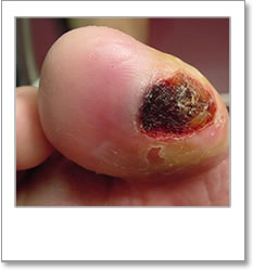
Active ulceration (5/16)
Active ulceration (pictures below) is defined by The International Working Group on the Diabetic Foot 2005 (IWGDF), as:
‘a full thickness wound, i.e. a wound penetrating through the dermis, below the ankle in a diabetic patient, irrespective of duration’.
NICE guideline [NG19]: Diabetic foot problems: prevention and management: 1.5 Diabetic foot ulcer recommend the SINBAD (site, ischaemia, neuropathy, bacterial infection, area and depth) or the TEXAS scale is used to classify a diabetic foot ulcer.
If during the screening process you discover the patient has a foot ulcer the patient should be referred without delay for treatment/management by an experienced podiatrist who is part of a multidisciplinary foot team/service.

Structural abnormality of the foot (4/16)
Structural abnormality of the foot (pictures below) is defined as
‘A change in foot shape that resulted in a difficulty in fitting shoes which could be purchased in high street shops’. (Scottish Diabetes Group – Foot Action Group 2010).
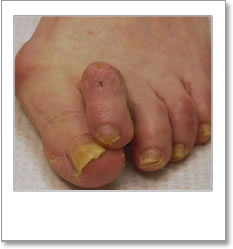


A non significant structural abnormality of the foot can be described as a very minor change of shape of the foot which does not result in areas of pressure, leading to callus formation, and a difficulty in fitting shoes which could be purchased in high street shops.
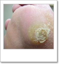
Significant callus (3/16)
Significant callus (pictures below) is defined as
‘Callus that requires Podiatric Management’ (Scottish Diabetes Group – Foot Action Group 2010).
Significant callus causes pressure on the underlying tissues which can result in the tissues breaking down and an ulcer developing. If a patient has significant callus and is not attending a podiatrist then they should be referred to have a treatment/management plan agreed and introduced to suit their needs.


Non significant callus can be described as callus that does not require podiatric treatment, does not pose any risk and can be treated/managed by the patient
The treatment of non significant callus or areas of dry skin can be managed by the patient after some simple instruction. The careful use of emery boards/ pumas stone, the regular application of a moisturiser cream and by following the advice given in the Low risk leaflet will usually achieve this.
Starting the screening process (2/16)
To start the screening process you should:
- Seat patient on examination couch/chair
- Inform the patient that you are going to examine their feet to check their circulation, sensation and any other risk factors that they might have which could lead to a foot problem related to their diabetes
- Request patient remove shoes and socks/stockings and assist if required
Ascertain the following:
- Has the person with diabetes been experiencing any problems with their feet since their last screening appointment?
- Has the person with diabetes noticed any changes to their feet since their last screening appointment?
- Is the person with diabetes complaining or any podatric-type problems (e.g. corns, calluses, nail problems, etc…)?
- Does the person with diabetes attend a podiatrist regularly?
04: The procedure (NHS England)
Aim and equipment (1/16)
Aim
The aim of carrying out a foot screening is to identify the presence of risk factors for diabetic foot complications which could lead to ulceration such as – Neuropathy, Peripheral Arterial Disease, Significant structural abnormalities, Significant callus, previous ulceration and the inability to self care.
Equipment
The only piece of equipment that is required to carry out a simple, evidence based, foot screening is a 10g monofilament. The monofilament used should be of good quality such as those manufactured by Bailey Instruments or Owen Mumford and should be used and replaced as per manufacturers instructions to ensure that the monofilament remains accurate. The length of time a monofilament will remain accurate will vary according to it’s frequency of use but Bailey Instruments and Owen Mumford recommend changing the monofilament after approximately 6 months of use. Many clinics use monofilaments much longer than this which can result in less accurate testing. The monafilament should always be replaced if bent.

Neurothesiometer
The Neurothesiometer is a device that tests a patient´s vibration perception threshold (VPT) and monitors diabetic neuropathy by measuring vibration sense. The device graduates in vibration intensity which ranges from 1 – 50 volts.
The Neurothesiometer is not recommended by NICE for use as part of the screening process, but can be useful as part of a more ‘in depth’ assessment in specialist centres.